Medical Imaging Services
Positron Emission Tomography (PET)
A positron emission tomography (PET) scan is an imaging test that helps measure how your tissues and organs
are functioning. A PET scan uses a radiopharmaceutical or tracer to show this activity. This scan can
provides a high degree of accuracy and precision and may sometimes detect disease before it shows up on
other types of imaging tests.
SPECT- Computed Tomography (SPECT-CT)
The most advanced form of CT scanning available and one of only 3 SPECT-CT machines in the Los Angeles area.
A SPECT-CT scan is a type of nuclear medicine scan where the images or pictures from two different types of
scans are combined together. The combined scan can provide precise information about how different parts of
the body are working and more clearly identify problems.
SPECT CT scans allow for images to be captured with the highest degree of precision when compared to
conventional cameras and allows for nuclear medicine services to be administered with the greatest level of
accuracy.
Nuclear Therapeutics
The administration of radiopharmaceuticals for diagnostic or treatment related purposes, nuclear
therapeutics involves the administration of radioisotopes as either an implant or injection. Your Oncologist
will work with you and the radiology team at TACRI to determine the most appropriate treatment course should
your clinical presentation meet the criteria to benefit from nuclear therapeutic services.
Radiofrequency Ablation (RFA)
Radiofrequency Ablation is a process that involves the use of a needle to travel underneath the skin to
nerve endings in an attempt to destroy nerves or clusters of cancerous cells connected to a particular area
of the body. This therapy is useful in the management of pain for cancer patients who require pain
management beyond what pharmaceuticals are able to offer and is an effective method for treating and even
removing cancer from the body.
Image guided radiofrequency ablation is an outpatient procedure and generally patients go home
the day of the procedure. These techniques have achieved marked success within the realm of oncology
including:
- The potential cure for liver metastases associated with metastatic colorectal cancer
- Lung cancer
- Renal cancer and adrenal gland cancer
- An alternative to more invasive surgical procedures
- Effective method for lasting chronic pain management that is non narcotic and relieving of the pain
associated with chemotherapy, radiation therapy and cancer itself.
-
Tumor tracking
Tumor tracking is a trend type measurement used to evaluate changes in tumors throughout the course of
therapy for oncology patients. Multiple diagnostic tools are available to provide the measurements
necessary to determine cancer progression. This allows for accurate, individualized and informed
decision making between you and your provider.
- Interventional and Vascular Radiology
Interventional radiology is a subspecialty of radiology where image-guided procedures are performed to
diagnose and treat diseases involving multiple organ systems and the vasculature. Using the least
invasive techniques currently available these procedures are precision based and have less risk, less
pain and less recovery time when compared to general surgery.
- Musculoskeletal imaging
Musculoskeletal imaging is a subspecialty of radiology involving the interpretation of images of bones,
joints and associated soft tissues and diagnosing injuries and disease.
- X-Ray
An X-ray is a quick, photograph-like test that produces images of the bones inside your body using small
amounts of radiation called X-rays. During the test, X-ray beams pass through your body, and they are
absorbed in different amounts depending on the material. X-ray imaging is able to allow for the
evaluation and interpretation of bones, fat, foreign objects and air within the body.
For some types of X-ray tests, a contrast medium — such as iodine or barium — may be administered to
provide a higher quality accuracy and detail.
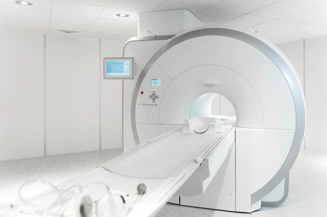
- MRI
Magnetic resonance imaging (MRI) is an imaging method which uses a magnetic field and radio waves to
build detailed images of the organs and tissues within your body.
MRI machines are commonly large tubes which hold very strong magnets. Patients lie down on a bench which
is then slid inside the tube, while inside the magnetic field temporarily moves water within your body
while the administration of radio waves allow for images to be taken and compiled into highly accurate
3D renderings of the inside of your body.
We are able to medicate and monitor patients with a history of claustrophobia and anxiety during the MRI
procedure when necessary.
- Ultrasound
Ultrasound is the generation of images by using the reflection of soundwaves off of the soft tissues
that make up your body. Ultrasound procedures are not painful, require little or no preparation and
involve the simple application of a gel on top of the area being examined.
Same day testing may be available and results of imaging may be ready in as little as one hour following
your appointment.